


I want to do home work for bio 120 you have to answer 34 questions
Question Description
Exercise 6 – DIFFUSION AND OSMOSIS
Student Learning Outcomes
At the completion of this exercise you should:
(l) Be able to define the terms diffusion and osmosis.
(2) Be able to list and discuss four mechanisms that cells use to move molecules across their plasma membranes.
(3) Be able to explain what Brownian motion tells us about atoms and molecules.
(4) Be able to explain the relationship between molecular weight and the rate of a molecules diffusion.
(5) Be able to list the characteristics of molecules that can, and those that cannot, move passively across a cells plasma membrane.
(6) Be able to describe how the solute concentration, inside of a cell, affects the rate (speed) of osmosis.
(7) Be able to define the following terms: concentration gradient, selectively permeable membrane, hypertonic, hypotonic, isotonic, and homeostasis.
-
Introduction
Virtually all life forms are composed of cells. The cell is called the fundamental unit of life because within it occur most of the biochemical life processes. One of the phenomena of life is that the chemical composition of a cell remains fairly constant, in spite of the fact that the cell continually uses substances from its external environment and at the same time discharges other substances into its environment: This state of chemical constancy in living systems is called homeostasis. This homeostasis, in addition to the fact that a cell’s surroundings are always of relatively different chemical composition from its inside, leads us to hypothesize that there must be some very selective means of chemical exchange across a cell’s plasma membrane. Today, we will investigate the processes of movement of some substances into and out of cells.
Question 1. Most “cells” do not appear to have an obvious “mouth” or other visible structures in their cell (“plasma”) membranes. Suggest one other way in which materials might be able to pass through the cell’s membrane:
|
Replace this text with your answer. |
Question 2. Cell biologists tell us that there are 4 basic mechanisms that cells use to get molecules across their membranes. You need to learn these 4 strategies. Go to your textbook, or other reference source, and define the following 4 mechanisms:
|
1.) |
Osmosis |
|
|
2.) |
Facilitated diffusion |
|
|
3.) |
Endocytosis and exocytosis |
|
|
4.) |
Active transport |
Brownian Motion
Robert Brown made an interesting observation in 1827 that led to the principle that “all atoms and molecules” are in constant motion. Dr. Brown was a botanist and army surgeon who was looking at particles inside pollen grains when he noticed the “rapid oscillatory motion of microscopic particles.” He later observed the same movement when looking at substances, like India Ink. India Ink is made of water and billions of suspended clumps of carbon atoms. Under high magnification, Brown observed that the clumps of carbon atoms were vibrating wildly in all directions. He hypothesized that moving water molecules, which cannot be seen, must be colliding with the clumps of carbon, forcing them to move. Further study has shown that Dr. Brown was correct and, today, we call this kind of observation “Brownian motion”.
A physical scientist would tell you that particles (atoms and molecules) are moving because “they have heat energy (Kinetic energy)”. If you ask what kinetic energy is, you will be told that it is “random molecular motion” which, of course, is a circular argument. The point here is that we really do not know the ultimate reason why all atoms and molecules on earth are moving, only that they are and that the more heat energy (“kinetic energy”) atoms or molecules have, the faster they move.
You are about to make this observation under the compound microscope yourself. The proper way to carry the compound microscope will be demonstrated. Always use two hands. Make sure that the cord is not dangling to prevent a tripping hazard. One hand should be holding the base of the microscope, while the other should hold the arm of the microscope.
Before using the microscope, check for the condition the microscope was left in from the prior class.
- Was the microscope placed back in its assigned compartment with the arm facing out toward you?
- Was the cord wrapped between the stage and objectives with the plug tucked inside the cord?
- Was the cord relatively untangled?
- Was the ocular lens clean? (If not, clean the dirty lens with the appropriate lens cleaner and lens paper (not a paper towel).
- Was the light turned off?
- Check that the Condenser is at its highest point, directly below the stage. There is a knob connected to the Condenser that allows it to be moved up and down.
- Was the mechanical stage centered so that the stage clips dont hang over the edge of the stage?
- Was the scanning objective lens (4x) (not another objective) placed over the stage? If not, rotate the nose piece until the scanning objective is facing the stage.
- Was the stage lowered to the lowest setting possible position? If not, use the coarse focus knob to do so, not the fine focus knob.
- Were there any slides remaining on the stage? If so, remove the slide and notify your instructor. It is important that the slide is placed in the correct box, or it may get lost.
- If any of these conditions were problematic, please let your instructor know.
Procedure:
- Plug in the cord and turn up the light intensity.to its maximum value and adjust the iris diaphragm to its most closed setting. As you proceed you can increase the light passing through the specimen by gradually opening the iris diaphragm.
- Adjust the distance between the oculars: Without placing the prepared slide on the stage yet, look through the oculars. You are likely going to see two circles of white light. Do not try to focus your eyes on any one thing, as nothing is in focus yet. Slowly, move the two oculars together and/or further apart until the two circles of white light become one circle of light, the Field of View.
- Lower the stage using the coarse focus knob, and make sure the (shortest) scanning objective is facing the stage, so that there is no chance of the slide scratching any objective lenses.
- Prepare your wet mount slide. Wash and dry a glass slide. Place a small drop of India ink on a clean slide. Place a cover slip on the slide.
- Coverslip: Carefully cover the preparation with a clean plastic coverslip as follows:
Place one edge of the coverslip near to the drop. The stain and water with which you mixed the cells will flow along the junction of the edge of the coverslip and the slide. Carefully lower the coverslip over the specimen keeping the edge of the coverslip in contact with the slide. In this way, the water will flow slowly and uniformly about the specimen and force out air bubbles from beneath the coverslip. (A few air bubbles are not a serious problem for your first slide.)

- Excess fluid: If liquid spills out or may spill out from under the coverslip, gently blot the excess with a towel, so that it will not later drip onto the stage of the microscope.
- Before looking through the eyepiece (ocular), open the stage clip, and place the slide on the stage of the microscope beneath the objective, with the coverslip visible on the upper side. The stage clip should be holding the slide in place, not pressing the slide under it. Using the left and right/up and down stage knobs), center the object below the objective without looking through the oculars. Nothing is in focus yet.
- Coarse adjustment with scanning lens: With the scanning lens in place, move the stage up to its highest point without looking through the oculars. Nothing is in focus yet.
Looking through the ocular with your right eye only (squint or cover your left eye), bring the specimen into focus by turning the coarse focus adjustment knob slowly until the specimen is generally in focus. Then turning the fine adjustment knob will bring the specimen into sharper focus.
- Focus your left eye: Viewing the specimen with both eyes through both oculars, turn the left ocular diopter until the specimen is clear in both eyes.
- Iris diaphragm: The light coming through the microscope may be either too bright or too dim. If the amount of light is not satisfactory, it can be adjusted by carefully regulating the size of the opening of the iris diaphragm by moving the lever beneath the stage. The iris diaphragm is part of the condenser which concentrates the light coming from the light source.
- Adjusting on low and high power objectives – use fine adjustment knobs only: If you wish to view the specimen using higher magnification, center the specimen in the field, and carefully rotate the revolving nosepiece to bring the next higher power objective into place beneath the body tube. The specimen will no longer be in focus. In order to sharpen the image of the specimen, adjust the focus using only the fine focus adjustment knob. (Again, the light may have to be adjusted with the iris diaphragm.) Each time you move to the next higher power objective, be sure you center the specimen beforehand.
- Examine the drop first under scanning, then low power, then under high power. Be sure you can see the individual particles of India ink.
- Focus your attention on one particle (under high power) for several seconds. Look for a slight but vigorous movement of this particle, independent of the other particles. (Note: You may need to wait a few minutes. Then, if you see a mass “flowing” movement of all the particles in one direction like a small river, this is not Brownian motion. Wait for the flowing to subside, then carefully observe one particle.)
Question 3. Describe the Brownian motion in your own words:
|
Replace this text with your answer. |
Question 4. Describe, in your own words, how this observation indicates that all visible and invisible molecules are in motion?
|
Replace this text with your answer. |
- Remove the slide: Once everyone in your group has viewed the Brownian Motion and you need to remove the slide, be sure to rotate the nosepiece to the scanning objective. Then using the coarse adjustment knob, lower the stage to its lowest position. Then open the stage clip and remove the slide.
Clean up.
- Rinse any wet mount slides and place them in the container marked Used slides. Wash and dry the coverslips and place them in their original container.
- Throw away any Kimwipes or other paper.
- You will need your microscope for other parts of this lab, so leave it available. However, make sure it is not near where there is sugar solution. Keep it safe.
II. DIFFUSION
Diffusion is the movement of particles from an area of high concentration to an area of low concentration. This results from the continuous random motion that is characteristic of all molecules in liquid or the gas states. A few observations about diffusion will help us to understand how molecules can move from one location to another, perhaps even across cell membranes.
A. Diffusion through a Colloid
The contents of a cell (the cytoplasm) may be described as a colloid rather than a liquid or solid. Large protein molecules are present in a cell’s cytoplasm that allow it to be in a transitional state of matter called a colloid (somewhere between a liquid and a solid). A special kind of colloid, agar gel, is available in the laboratory and will be used to demonstrate how molecules diffuse from one place to another once they are inside a cell.
Agar is a carbohydrate extracted from algae in powder form. A gel is prepared by mixing the powder with water, then heating followed by cooling–similar to the preparation of a gelatin (animal protein) dessert. The result is a gelatin-like substance composed of intertwined molecules with water trapped among them. Two compounds (molecules), potassium permanganate and methylene blue, have been selected to illustrate diffusion through a colloid. Unlike most components of living cells, these compounds are brightly colored, allowing us to watch their diffusion.
Procedure:
1. Working with your team, obtain a disposable Petri dish containing agar gel.
2. Using a No. 5 cork borer (a “punch”), make two holes in the agar approximately 5 centimeters apart (See diagram below). Remove the plugs of agar with a toothpick and place in the trash.
3. Bring your agar dish, the dropper bottles of potassium permanganate solution and the dropper bottle of methylene blue back to your lab bench.
4. Place two drops of 1% potassium permanganate solution in one of the holes. In the other hole, place two drops of 1% methylene blue solution. Start a timer for 60 minutes. Return the bottles back to the side lab benches for others to use.
5. After approximately one hour, measure the diameter (in millimeters) of the circle that the solutions diffused.
|
Potassium permanganate: |
mm |
Methylene blue: |
mm |
6. Examine your Petri dish. Shade the agar in the diagram to demonstrate the movement of the solutions in the diagram below. Shade to show where the concentration of the potassium permanganate is higher and where it is lower.
Top view of Petri dish: Side view of Petri dish:
of the agar cut out. Represented by a circle with two smaller circles inside
Right:
Side view of Petri dish with two sections of the agar cut out.” src=”https://docs.google.com/drawings/u/0/d/sw001XxjnioFkNL8IWlK6NA/image?w=428&h=163&rev=1&ac=1&parent=1SUAcijEhcOpgkeRbRil_eOcx-8Rgk2_BZhOOaDXfR_M”>
Question 5. Your Petri dish illustrates the most basic characteristic of diffusion. Complete the following to understand this basic principle:
The net diffusion of potassium permanganate appears to be from the area of its ___ concentration to the area of its _________ concentration.
(“lower to higher” – or – “higher to lower”?)
Question 6. In addition to concentration, the molecular weight of a diffusing molecule is also an important factor that can determine the rate (speed) of diffusion. We happen to know that potassium permanganate’s molecular weight is 150, while that of a methylene blue molecule is 374.
Use your own observations (above) to explain how molecular weight appears to affect the rate of diffusion here:
|
Replace this text with your answer. |
Question 7. Based on the results, what is the relationship between molecular weight and diffusion rate?
Highlight one: Inverse Direct
State the basis of your conclusion:
|
Replace this text with your answer. |
Question 8. How would you expect the dyes to be distributed in the agar gel after several days? Would you expect to still see evidence of a concentration gradient?
|
Replace this text with your answer. |
Question 9. Calculate the molecular weights for the following molecules so you can predict which will diffuse more rapidly.
Note: Molecular weights are calculated by adding up the atomic weights (masses) for all the atoms that make up a given molecule.)Look at the Periodic Table on the wall in the laboratory. It will give you the atomic weights of all the atoms in the two molecules below.
Calculate the weight of these two molecules. Show your work here:
|
Water, H2O |
Sucrose, C12H22O11 |
|||||
|
Number |
At. mass |
Total |
Number |
At. mass |
Total |
|
|
H: |
||||||
|
O: |
||||||
|
C: |
||||||
|
Total: |
Total: |
|||||
Question 10. Which substance, water or sucrose, is more likely to be able to diffuse through a semipermeable membrane, and on what basis did you make your decision?
|
Replace this text with your answer. |
Clean up.
1. Throw away your Petri dish, agar included, into the trash can.
2. Wipe down all countertops and student benches with yellow cleaning solution.
III. LIVING CELL MEMBRANES
The membranes that surround all cells allow some molecules to diffuse across while inhibiting others. In other words, the membranes of cells are said to be “selectively permeable.” The membranes of all cells are made out of 2 layers of phospholipid molecules with various kinds of channels passing through them. (See Figure 1). Phospholipids are each made of the phosphate-containing head which is hydrophilic, and two long fatty acid chains which are hydrophobic.
Question 11. Phospholipids are each made of the phosphate-containing head which is hydrophilic (easily combine with water), and two long fatty acid chains which are hydrophobic (do not combine with water, fatty acids are oils). Color the circular phosphate heads blue; color the fatty acid chains of the phospholipids red. Then label them hydrophobic and hydrophilic based on your reading above.
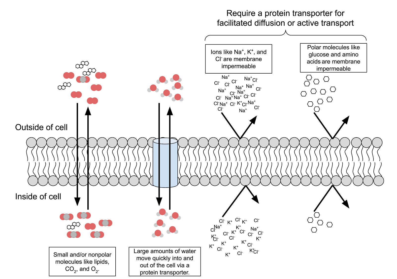
Figure 1. Selectively Permeable Cell Membrane
Question 12. Observe Figure 1.What kinds of molecules are able to pass through the phospholipid bilayer membrane easily? What kinds of molecules are not able to pass through easily? What are the characteristics of each (charge, polar vs. nonpolar), and size?
|
Move easily |
Do not move easily |
Because water is a small and flat molecule (despite it being polar), it can either pass through the phospholipid bilayer membrane. It can more quickly pass into and out of cells through tunnel-like proteins called aquaporins that traverse the cell membrane.
Question 13. Label the aquaporin protein in Figure 1. What does the aquaporin allow to pass through the membrane?
|
Replace this text with your answer. |
IV. Osmosis and the rate (speed) of diffusion along a concentration gradient
Often, scientists find it helpful to construct a simplified model of an object or phenomenon in order to understand it more clearly. This is exactly what you will do now. You will make several different model cells using a plastic membrane material (dialysis tubing) that mimics the osmosis characteristics of a living cell’s membrane.
The speed or rate at which a molecule diffuses from one area to another depends on the concentration gradient between the two areas. For example, if the concentration of perfume molecules is higher in one room compared to an adjacent room connected by an open door, we would say the concentration gradient between the two rooms is very steep and the rate of diffusion would be very rapid. Conversely, if the concentration of perfume molecules was equal in the two rooms, then the rate of diffusion would be zero and the net movement of the perfume molecules would stop. (Movement would still occur, as Brownian Motion still occurs, but the diffusion would be at equal rates between the rooms.)
Osmosis follows the same laws as diffusion but always refers to water, the principle solvent in cells. A solvent is a fluid that dissolves substances, while the term solute is used to describe substances dissolved in a solvent to make a solution. This means that water will move down its concentration gradient, too, from an area of high concentration of water to an area of low concentration of water.
Because water is the universal solvent in all cells its diffusion into or out of cells is critical in living organisms. Too much water entering or leaving a cell will cause cell death and often the death of the entire organism. You will now set up several different model cells and measure the direction and rate (speed) of osmosis.
Question 14. Define the terms Solvent and Solute:
|
Replace this text with your answer. |
Question 15. Below is a diagram of three model cells filled with three different solutions. Make a prediction. Review Figure 2 and complete Table 1 to its right. For column 4, predict whether the model cell will increase or decrease in weight after one hour. (Hint: Will the net diffusion of water be into the model cell or into the beaker?)
Baggy B (representing Cell B) contains 10 mL of 25% sucrose.
Baggy C (representing Cell C) contains 10 mL of 50% sucrose.
The beaker contains DI water (0% solutes).” src=”https://docs.google.com/drawings/u/0/d/sncrbRTGdNJPLGQF5jiTung/image?w=283&h=375&rev=2&ac=1&parent=1SUAcijEhcOpgkeRbRil_eOcx-8Rgk2_BZhOOaDXfR_M”>
Table 1: Prediction for Model Cells
|
Cell Letter |
% solutes in cell |
% water in cell |
Increase or decrease after 1 hour? |
|
A |
|||
|
B |
|||
|
C |
Figure 2. Diagram showing the experimental set up for the 3 model cells
Procedure:
- Obtain a three pieces of paper towel on which to place your plastic tubing. This tubing is like plastic wrap or plastic bags. While unseen by the naked eye, the plastic has molecular holes of a specific size that allow some molecules to diffuse across the plastic while blocking other, larger, molecules. Therefore, the plastic tubing is selectively permeable. This tubing will simulate the structure of the membrane
- Obtain a short stack of paper towels, wet the center towels in the stack with deionized water, so you will have dry towels on the bottom and top and wet towels in the middle. Use this bed to keep you plastic tubing moist. Allowing the tubing to dry out will cause it to lose its permeability properties
- Obtain a beaker, and fill it 2/3 full with deionized water
- Obtain 3 pieces of water-soaked plastic tubing 20 cm long and six pieces of thread. Twist one end of the first plastic tubing, and fold down. Then tie the folded end tightly with a double knot.
5. Open the other end of the tube by rolling with your fingers. Dont let the tubing get dry, or it may crack.
6. Write down the colors of the solutions here:
|
1% |
25% |
50% |
7. Fill the 3 plastic tubes with the contents shown below in Figure 2.
8. Twist the other end of the plastic tubing, then fold down and tie a string around that end. The baggie you have created should not be entirely full of water, but instead have a bit of space remaining at the top. Tie the folded ends securely. The goal is to create a water-tight bag.
9. Check for leaks. First, press any extra liquid out of the ends created by filling the bags. Then, by gently but firmly rolling the bag on the paper towel, checking for leaks. Then trim the excess thread. Keep the model cells on the clean paper towel between measurements of cells. Have your instructor check.
10. Check to see if your Triple Beam balance is calibrated properly This means the line on the end of the center beam lines up at the zero line when all of the weights (poises) are slid to the left at zero. If the lines do not meet at the zero line, ask your instructor to assist you.
- Next, blot excess water off the model cells, and place it on the center of the balance pan. First, move the heaviest weight to the right to the first notch which causes the pointer to drop, then, move it back one notch, causing the pointer to rise. If the pointer goes below the zero, the weight is too much, so you will need to try a lighter weight. If it stays above the zero, you will need to add more weight.
- Add weights as necessary, moving along one beam and then the next lighter beam until the pointer goes to zero. The weight of the specimen is the sum of the values for all of the weight positions, read directly from the graduated beams.
- Weigh each model cell to nearest 0.01 grams using the balance on your lab bench. If you are not sure how to read this measurement on the Triple Beam balance, please ask your instructor to assist you. Record the “cell” weights in Table 2 in the column titled “initial weight.”
14. Place cells A, B, and C simultaneously in a beaker filled with 100% distilled water (solvent) (See Figure 2 again).
|
Note the time here: |
15. Remove all 3 model cells from their beaker every 15 minutes for the next hour. Place the model cells in your paper towel bed. Blot dry and weigh each model cell, being careful to dry the string and the area where you have folded the tubing. Weigh the cells again to the nearest 0.1 gram. Handle the cells very carefully to avoid causing leaks. After you have weighed and recorded the weight, return the “cells” back to the beaker for the next 15-minute period
16. At 15-minute intervals, for 1 hour, record the “total weight” of each cell in Table 2.
17. Calculate the net mass change for each model cell in the last row of Table 2.
Net mass change = Final mass (at time 60) initial mass (at time 0)
Table 2. Change in Mass of Three Model Cells by Osmosis over Time
|
Mass (g) |
|||
|
Time (minutes) |
Cell A |
Cell B |
Cell C |
|
0 |
|||
|
15 |
|||
|
30 |
|||
|
45 |
|||
|
60 |
|||
|
Net mass change |
|||
Question 16. How do your results compare with your prediction in Table 1?
|
Replace this text with your answer. |
Graph the Osmosis data
1. Read all of the following directions and use google sheets, excel or some other program to graph your data as a line graph. Label the graph with the vertical (“Y”) axis as “Mass (grams)” and the horizontal (“X”) axis as “Time (minutes).
2. Plot the data from Table 2 for “Mass” at each of the 15-minute intervals.
- Include the data for the three model cells as 3 separate curves (lines) using three different colors on the same graph. Create a key for identifying the colored line according to its corresponding Cell letter and % Solutes.
- Write the title Change in Mass of Three Model Cells with Varying Concentration of Solutes via Osmosis over Time at the top of the graph.
|
Replace this text with your graph |
Clean up
- Drain the sugar water from the plastic tubing down the sinks, and then rinse off the tubing. Flush the sink with plenty of water. Throw the plastic tubing in the trash.
- Spray a clean paper towel with the yellow cleaning solution, and then wipe your Triple Beam balance pan.
- Wipe off all surfaces, including your lab bench, the side lab benches (under the sugar solution dispersal containers). (Sugar attracts ants!)
- Throw all paper towels away.
Hypotonic, Hypertonic, and Isotonic Environments.
The three terms above are used to describe solute concentration environments a cell may find itself in. In two of these environments water will enter or leave the cell and the cell will change shape and perhaps die. You must learn what these terms mean so you are able to answer questions that use them.
These three terms always describe the solute concentration on one side of cell’s membrane relative to the solute concentration on the other side of the cell membrane.
The term hypotonic means “less solute concentration here” relative to the other side of a cell’s membrane. If you are told the outside environment a cell finds itself in is hypotonic, then you automatically know that the inside environment is the exact opposite or hypertonic.
As you recall, the cell membrane will not allow large or ionic substances to move across it, but water may pass. Your job will always be to determine whether the cell will swell up with water or shrink due to water loss. To do this you must decide which side of the membrane has a higher water concentration. Once this is done you can predict which way water will move (diffuse) since molecules (such as water) always diffuse from an area of higher concentration to an area of their lower concentration.
Look at the osmosis data you collected for Model cell A. We had you put a 1% solute (sucrose) concentration inside the cell and surrounded it with deionized water (a solution that had 0% solute concentration). The proper terminology would be that the inside of the model cell was hypertonic (greater solute concentration) compared to the outside environment that would be described as
Have a similar assignment? "Place an order for your assignment and have exceptional work written by our team of experts, guaranteeing you A results."

Our Service Charter
1. Professional & Expert Writers: Essay Noon only hires the best. Our writers are specially selected and recruited, after which they undergo further training to perfect their skills for specialization purposes. Moreover, our writers are holders of masters and Ph.D. degrees. They have impressive academic records, besides being native English speakers.
2. Top Quality Papers: Our customers are always guaranteed of papers that exceed their expectations. All our writers have +5 years of experience. This implies that all papers are written by individuals who are experts in their fields. In addition, the quality team reviews all the papers before sending them to the customers.
3. Plagiarism-Free Papers: All papers provided by Essay Noon are written from scratch. Appropriate referencing and citation of key information are followed. Plagiarism checkers are used by the Quality assurance team and our editors just to double-check that there are no instances of plagiarism.
4. Timely Delivery: Time wasted is equivalent to a failed dedication and commitment. Essay Noon are known for the timely delivery of any pending customer orders. Customers are well informed of the progress of their papers to ensure they keep track of what the writer is providing before the final draft is sent for grading.
5. Affordable Prices: Our prices are fairly structured to fit in all groups. Any customer willing to place their assignments with us can do so at very affordable prices. In addition, our customers enjoy regular discounts and bonuses.
6. 24/7 Customer Support: At Essay Noon, we have put in place a team of experts who answer all customer inquiries promptly. The best part is the ever-availability of the team. Customers can make inquiries anytime.